
ファイル:Echinococcus Life Cycle.svg

この SVG ファイルのこの PNG プレビューのサイズ: 629 × 600 ピクセル. その他の解像度: 252 × 240 ピクセル | 504 × 480 ピクセル | 806 × 768 ピクセル | 1,074 × 1,024 ピクセル | 2,149 × 2,048 ピクセル | 1,280 × 1,220 ピクセル。
元のファイル (SVG ファイル、1,280 × 1,220 ピクセル、ファイルサイズ: 643キロバイト)
ウィキメディア・コモンズのファイルページにある説明を、以下に表示します。
|
概要
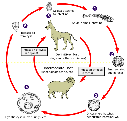
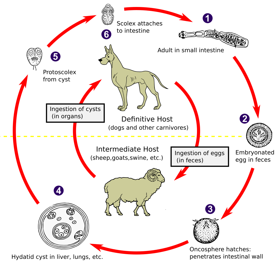
| 解説 | The adult Echinococcus granulosus (3 to 6 mm long) [1] resides in the small bowel of the definitive hosts (dogs or other carnivores). Gravid proglottids release eggs [2] that are passed in the feces. After ingestion by a suitable intermediate host (under natural conditions: sheep, goat, swine, cattle, horses, camel), the egg hatches in the small bowel and releases an oncosphere [3] that penetrates the intestinal wall and migrates through the circulatory system into various organs, especially the liver and lungs. In these organs, the oncosphere develops into a cyst [4] that enlarges gradually, producing protoscolices and daughter cysts that fill the cyst interior. The definitive host becomes infected by ingesting the cyst-containing organs of the infected intermediate host. After ingestion, the protoscolices [5] evaginate, attach to the intestinal mucosa [6] and develop into adult stages [1] in 32 to 80 days. The same life cycle occurs with E. multilocularis (1.2 to 3.7 mm), with the following differences: the definitive hosts are foxes, and to a lesser extent dogs, cats, coyotes and wolves; the intermediate host are small rodents; and larval growth (in the liver) remains indefinitely in the proliferative stage, resulting in invasion of the surrounding tissues. With E. vogeli (up to 5.6 mm long), the definitive hosts are bush dogs and dogs; the intermediate hosts are rodents; and the larval stage (in the liver, lungs and other organs) develops both externally and internally, resulting in multiple vesicles. E. oligarthrus (up to 2.9 mm long) has a life cycle that involves wild felids as definitive hosts and rodents as intermediate hosts. Humans become infected by ingesting eggs , with resulting release of oncospheres in the intestine and the development of cysts in various organs. Image adapted from original available at the United States Centres for Disease Control Parasitology Identification Laboratory ([1]). |
| 日付 | |
| 原典 |
このファイルの派生元: Echinococcus Life Cycle.png: |
| 作者 |
CDC ベクタ: 🎱 |
| SVG 開発 |
ライセンス
この画像は、アメリカ合衆国保健福祉省の一部である疾病予防管理センターの著作物であり、職員の公務の一環として撮影または作成されたものです。アメリカ合衆国連邦政府の著作物として、画像はパブリックドメインの状態にあります。
|
元のアップロードログ
This image is a derivative work of the following images:
- Echinococcus Life Cycle.png licensed with PD-USGov-HHS-CDC
- 2007-01-24T10:54:56Z Pngbot 600x571 (45555 Bytes) optimized with optipng
- 2005-04-26T01:48:50Z FirstPrinciples~commonswiki 600x571 (55999 Bytes) Smaller & clearer
- 2005-04-26T01:36:23Z FirstPrinciples~commonswiki 800x761 (80990 Bytes)
Uploaded with derivativeFX
ファイルの履歴
過去の版のファイルを表示するには、その版の日時をクリックしてください。
| 日付と時刻 | サムネイル | 寸法 | 利用者 | コメント | |
|---|---|---|---|---|---|
| 現在の版 | 2021年2月1日 (月) 01:31 | | 1,280 × 1,220 (643キロバイト) | Pixelsquid | Resized. |
| 2021年1月31日 (日) 20:44 |  | 320 × 305 (460キロバイト) | Pixelsquid | == {{int:filedesc}} == {{Information |Description=The adult Echinococcus granulosus (3 to 6 mm long) [1] resides in the small bowel of the definitive hosts (dogs or other carnivores). Gravid proglottids release eggs [2] that are passed in the feces. After ingestion by a suitable intermediate host (under natural conditions: sheep, goat, swine, cattle, horses, camel), the egg hatches in the small bowel and releases an oncosphere [3] that penetrates the intestinal wall and migrates through the... |
ファイルの使用状況
グローバルなファイル使用状況
以下に挙げる他のウィキがこの画像を使っています:
- ar.wikipedia.org での使用状況
- arz.wikipedia.org での使用状況
- be.wikipedia.org での使用状況
- bs.wikipedia.org での使用状況
- ca.wikipedia.org での使用状況
- dag.wikipedia.org での使用状況
- el.wikipedia.org での使用状況
- en.wikipedia.org での使用状況
- es.wikipedia.org での使用状況
- fa.wikipedia.org での使用状況
- ga.wikipedia.org での使用状況
- gl.wikipedia.org での使用状況
- hi.wikipedia.org での使用状況
- hu.wikipedia.org での使用状況
- hy.wikipedia.org での使用状況
- ia.wikipedia.org での使用状況
- id.wikipedia.org での使用状況
- is.wikipedia.org での使用状況
- it.wikipedia.org での使用状況
- ko.wikipedia.org での使用状況
- ky.wikipedia.org での使用状況
- lt.wikipedia.org での使用状況
- mk.wikipedia.org での使用状況
- ml.wikipedia.org での使用状況
- ms.wikipedia.org での使用状況
- nl.wikipedia.org での使用状況
- om.wikipedia.org での使用状況
- or.wikipedia.org での使用状況
- pl.wikipedia.org での使用状況
- pt.wikipedia.org での使用状況
- ro.wikipedia.org での使用状況
- ru.wikipedia.org での使用状況
- sl.wikipedia.org での使用状況
- sr.wikipedia.org での使用状況
- sv.wikipedia.org での使用状況
- th.wikipedia.org での使用状況
- tl.wikipedia.org での使用状況
- tr.wikipedia.org での使用状況
- uk.wikipedia.org での使用状況
- uz.wikipedia.org での使用状況
- vi.wikipedia.org での使用状況
- www.wikidata.org での使用状況
- yo.wikipedia.org での使用状況
- zh.wikipedia.org での使用状況